All About Glaucoma (Updated 2026)
Introduction
Glaucoma remains the leading cause of irreversible blindness worldwide, affecting more than 70 million individuals, of whom approximately 10% are bilaterally visually impaired. The disease often progresses gradually and may remain asymptomatic until advanced stages, resulting in a substantial proportion of undiagnosed cases across the world. Consequently, early detection and prompt intervention are paramount to preventing irreversible visual impairment.
Pathophysiologically, glaucoma encompasses a group of progressive optic neuropathies characterized by retinal ganglion cell (RGC) loss, optic nerve head excavation, and corresponding visual field defects. It is broadly classified into two primary subtypes: Primary Open-Angle Glaucoma (POAG) and Primary Closed-Angle Glaucoma (PCAG). Secondary glaucomas may arise secondary to ocular trauma, chronic corticosteroid use, intraocular inflammation, neoplastic processes, or other etiologies such as pigment dispersion syndrome and pseudoexfoliation.
Risk factors
Primary Open-Angle Glaucoma (POAG)
Primary Open-Angle Glaucoma is more likely to occur in individuals with a positive family history, those of Black race, or older adults. Primary care physicians play a crucial role in early detection by recognizing patients at risk, including those receiving systemic or topical corticosteroids, and by referring patients with a family history of glaucoma for a comprehensive ophthalmologic examination. Any individual with a family history of glaucoma who has not undergone a fundus examination in the past two years should be referred for glaucoma assessment.
Intraocular pressure (IOP) is closely associated with retinal ganglion cell loss, which exerts mechanical stress on the posterior structures of the eye. Elevated IOP arises from an imbalance between aqueous humor production by the ciliary body and its drainage through the trabecular meshwork and uveoscleral pathways. In open-angle glaucoma, elevated IOP occurs when there is increased resistance to aqueous outflow at the trabecular meshwork, whereas in angle-closure glaucoma, access to the drainage pathways is obstructed. Although glaucomatous optic neuropathy can develop in individuals with IOP within the normal range, elevated IOP remains the most significant modifiable risk factor.
Primary Closed-Angle Glaucoma (PCAG)
PCAG differs from POAG mainly in that the anterior chamber angle, the site where aqueous humor drains,is obstructed by abnormalities of the iris or lens, resulting in anatomical angle closure. The most common underlying mechanism is a pupillary block, where resistance to aqueous humor flow from the posterior to anterior chambers at the pupil leads to fluid accumulation behind the iris. This increases the iris convexity and causes the angle to close. PCAG can also occur due to dynamic physiological factors such as increased iris volume during pupil dilation or choroidal effusion.
In fewer than one-third of cases, patients with PCAG may present with an acute attack of primary angle closure. This condition manifests with pronounced conjunctival redness, corneal edema, a fixed mid-dilated pupil, shallow anterior chamber, and markedly elevated intraocular pressure. Affected individuals often experience ocular pain, nausea, vomiting, and transient blurring of vision with haloes around lights.
Risk factors include female sex, advancing age, and Asian ethnicity. Ocular risk factors primarily involve a crowded anterior segment in smaller eyes, characterized by a shallow central anterior chamber, a thicker and more anteriorly positioned lens, and a shorter axial length.
Clinical Presentation and Diagnosis

Tonometry
Intraocular pressure (IOP) can be measured using two primary methods: non-contact tonometry and contact tonometry. Non-contact tonometry uses a gentle puff of air to flatten the cornea, allowing the device to estimate the IOP without touching the eye. It is quick, non-invasive, and commonly used for screening purposes in clinics. Contact tonometry is a technique involving direct contact with the cornea, usually after applying a topical anesthetic. The most widely used form is Goldmann applanation tonometry, considered the gold standard for IOP measurement due to its high accuracy. Other types include Tono-Pen and rebound tonometers, which offer portable or handheld options. Both methods have established normal ranges, typically 10–21 mmHg, although this can vary slightly depending on the device and patient-specific factors such as corneal thickness.
Gonioscopy
The distinctive clinical features of angle closure are observed in the angle of the eye by gonioscopy. A simple, handheld, mirrored instrument is placed on the patient’s eye, followed by examination of the angle using a slit-lamp biomicroscopy. With indentation, the examiner is also able to determine if peripheral anterior synechiae (adhesions between the iris and trabecular meshwork) are present. Gonioscopy is highly subjective, with poor reproducibility, and gonioscopic findings may vary with the amount of light used during the examination or mechanical compression of the eye.
Ophthalmoscope, Slit-lamp fundus biomicroscopy and fundus photography
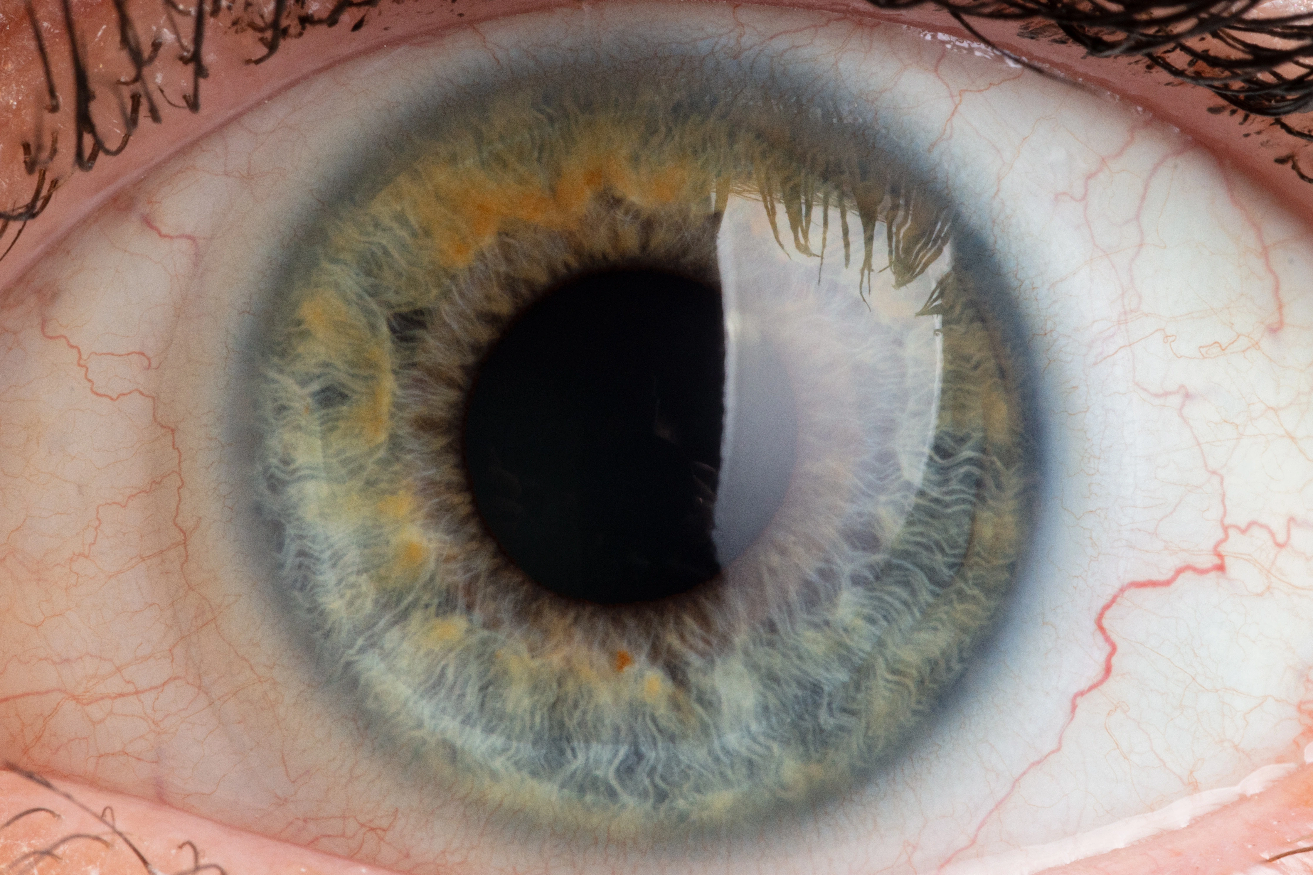
In patients with glaucoma, ocular examination typically reveals an increased cup-to-disc ratio, asymmetry in cup-to-disc measurements between the eyes, bayonetting retinal vessels, thinning of optic nerve tissues or disc hemorrhage. These findings can be assessed through direct observation of the optic nerve head using an ophthalmoscope or by indirect fundus examination via slit-lamp fundus biomicroscopy or optic nerve head photographs. Dilated fundus examination of the optic nerve head remains crucial for early detection as it may prompt for further assessment to confirm the diagnosis.
Optical Coherence Tomography (OCT) and Humphrey Visual Field (HVF)
Loss of retinal ganglion cells and optic nerve fibers results in characteristic changes in the optic nerve head and retinal nerve fiber layer, which can be identified on examination with the use of advanced imaging techniques, such as OCT, which provides objective and quantitative information regarding the extent of optic nerve fiber loss. Progressive retinal ganglion cell loss leads to deterioration of the visual field, typically starting in the midperiphery and advancing centripetally until only central or peripheral vision remains which can be detected by the HVF test. Although the presence of characteristic visual field defects can confirm the diagnosis, standard visual field testing may not detect abnormalities until 30–50% of retinal ganglion cells have already been lost. Visual field testing is still essential in monitoring the progression of the condition by comparison of the remaining functional visual field.
Treatment strategies
Medication
The primary objective in glaucoma management is to slow disease progression and maintain the patient’s quality of life. According to the American Academy of Ophthalmology Preferred Practice Pattern guidelines, treatment aims to lower IOP to a target level — defined as the pressure at which the clinician believes progression will be sufficiently reduced to prevent functional vision loss. This target IOP should be achieved with the fewest medications possible and minimal adverse effects.
Prostaglandin analogues (Latanoprost, Travoprost, and Bimatoprost) are generally the first-line agents. They enhance aqueous humor outflow through the uveoscleral pathway, are administered once nightly, and have few systemic side effects. However, local adverse effects may occur, including conjunctival redness, elongation and darkening of eyelashes, orbital fat atrophy, iris pigmentation, and periocular skin darkening.
Second-line topical medications are less effective in lowering IOP but may be used when prostaglandin analogues are contraindicated or not well tolerated. These include β-blockers (Timolol and Betaxolol), α-adrenergic agonists (Brimonidine), carbonic anhydrase inhibitors (Dorzolamide, Brinzolamide, and Acetazolamide), and cholinergic agents (Pilocarpine), which can be used individually or in combination. Prostaglandin analogues and carbonic anhydrase inhibitors are effective throughout the day and night, while β-blockers and α-adrenergic agonists primarily act during daytime.
Certain agents, particularly β-adrenergic blockers, may cause systemic side effects and are contraindicated in patients with chronic obstructive pulmonary disease, asthma, or bradycardia. To minimize systemic absorption of topical medications, patients should be advised to perform gentle punctal occlusion or keep their eyes closed for two minutes after application. Treatment success depends greatly on patient adherence, and reinforcing the importance of compliance is essential for optimal long-term outcomes.
Laser therapy
When medical therapy fails to achieve sufficient IOP reduction or causes unacceptable side effects, laser or incisional surgical intervention may be indicated. In POAG, laser trabeculoplasty can be performed to enhance aqueous humor outflow through the trabecular meshwork, thereby lowering IOP. The procedure is safe, minimally invasive, and typically performed in an outpatient setting.
The first-line treatment for PACG is laser peripheral iridotomy, which involves creating a full-thickness opening in the peripheral iris to relieve pupillary block and facilitate aqueous humor flow. This procedure is generally painless, straightforward and well tolerated by most patients.
In cases of acute angle-closure attack, the immediate goal is to rapidly reduce IOP using topical and systemic medications to minimize optic nerve damage. Once IOP is stabilized, laser peripheral iridotomy is then performed to relieve pupillary block and terminate the attack. Prompt and adequate pressure control often results in full visual recovery without permanent optic disc or visual field damage.
If conventional medical therapy fails to control an acute attack or is not tolerated, laser iridoplasty, which contracts the peripheral iris to widen the angle, may be performed. In situations where iridotomy is unsuccessful, surgical iridectomy is indicated.
For individuals at risk of PACG, particularly those with a family history of angle closure or symptoms of intermittent attacks, prophylactic iridotomy may be recommended to prevent irreversible trabecular meshwork damage and glaucomatous optic neuropathy.
Surgical options
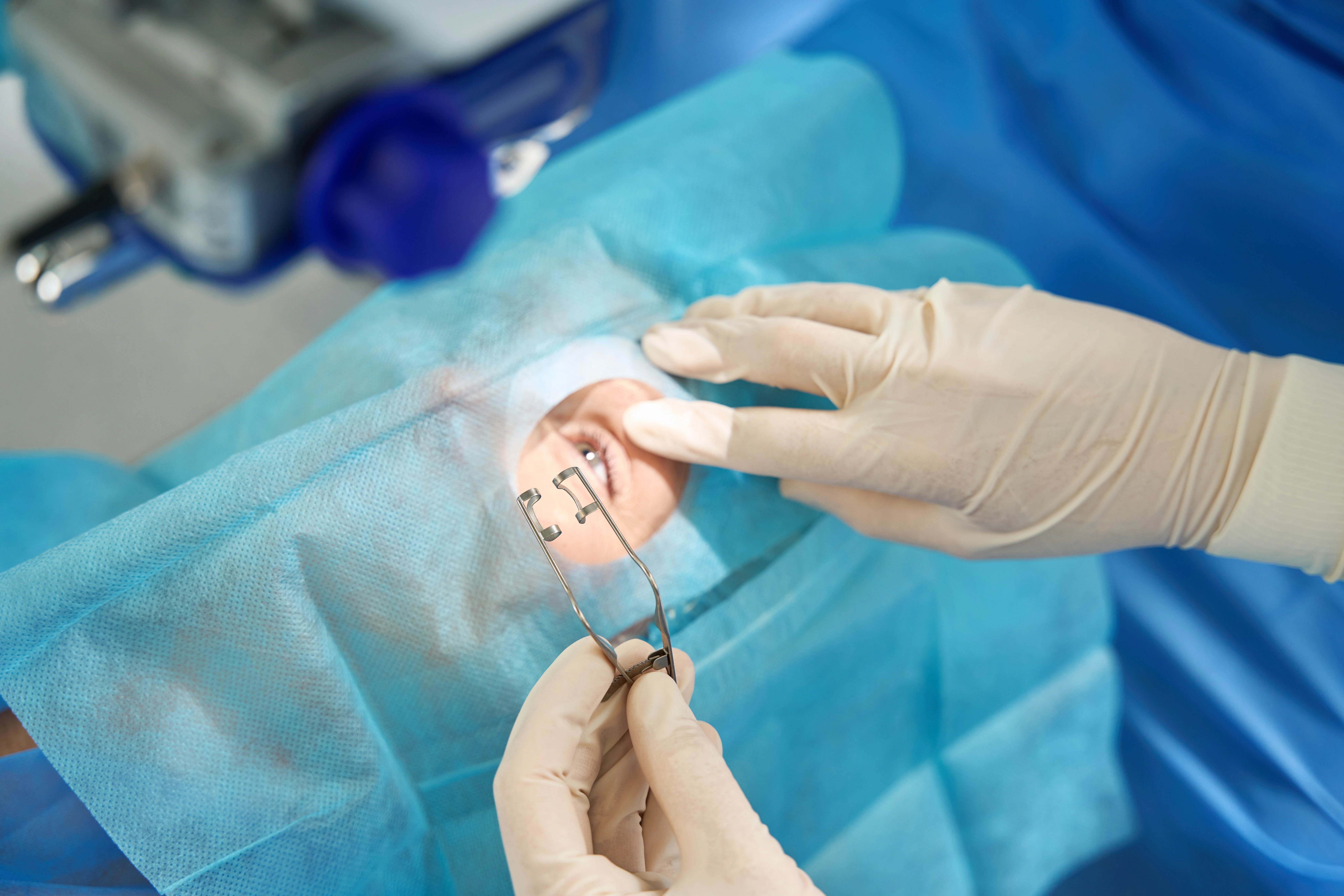
In patients with poor treatment adherence or advanced disease, surgical intervention may be considered as first-line therapy. Trabeculectomy remains the standard incisional surgery for lowering IOP. It involves excision of a small section of the trabecular meshwork and adjacent corneoscleral tissue, creating a drainage pathway for aqueous humor to exit the anterior chamber into the subconjunctival space for absorption.
Similar to POAG, trabeculectomy is indicated when IOP remains uncontrolled or progressive optic nerve or visual field damage occurs despite optimal medical and laser therapy. In patients with significant cataract, cataract extraction with intraocular lens implantation serves as an effective alternative, as it can reduce IOP, deepen the anterior chamber angle, and improve visual acuity.
In conjunction with cataract surgery, a minimally invasive glaucoma surgery (MIGS) procedure such as iStent implantation may also be performed. This involves inserting a microscopic stent into the trabecular meshwork to enhance aqueous outflow. The procedure is safe and typically recommended for patients with mild to moderate glaucoma, particularly those who experience difficulty adhering to topical medication regimens.
For more advanced cases where other glaucoma treatments have failed or are unlikely to succeed, glaucoma drainage implants (also known as tube shunts) may be employed to achieve long-term IOP control. These devices consist of a small tube inserted into the anterior chamber and a plate positioned beneath the conjunctiva, providing an alternative drainage pathway for aqueous humor outflow from the eye.
Conclusion
Glaucoma is a chronic, multifactorial optic neuropathy where early diagnosis and pressure control are key to preserving vision. Primary care and eye care professionals play vital roles in identifying high risk patients, ensuring adherence to therapy and monitoring for progression and adverse effects.
Summary
| Type of Treatment | Examples / Procedures | How It Works | When It’s Used | Things to Know |
| Eye Drops (Medications) | Prostaglandin analogues (Latanoprost, Travoprost, Bimatoprost) | Helps drain fluid from the eye to lower eye pressure | Usually the first treatment option | Used once daily at night; may cause mild redness, longer eyelashes, or darkening of the iris/skin |
| Beta-blockers (Timolol, Betaxolol) | Reduces the amount of fluid the eye produces | Used if first-line drops are not enough | May affect heart and lungs – not suitable for people with asthma or heart problems | |
| Alpha-agonists (Brimonidine) | Lowers eye pressure by reducing fluid production and increasing drainage | Used together with other drops | May cause dry mouth or tiredness | |
| Carbonic anhydrase inhibitors | (Dorzolamide, Brinzolamide) | Reduces fluid production inside the eye | Used alone or with other drops | May cause mild stinging or a bitter taste |
| Cholinergic agents (Pilocarpine) | Opens the eye’s drainage channels | Sometimes used in angle closure glaucoma | Can cause small pupils or blurry vision, especially at night | |
| Laser Treatments | Laser Trabeculoplasty (for POAG) | Improves drainage through the eye’s natural filter | When drops don’t lower pressure enough | Quick and safe clinic procedure; may need repeat treatments |
| Laser Peripheral Iridotomy (for PACG | Creates a small hole in the iris to improve fluid flow | First-line treatment for angle closure glaucoma | Painless clinic procedure; usually done once | |
| Laser Iridoplasty | Shrinks part of the iris to open the angle | Used when other laser treatment isn’t enough | Quick and well tolerated | |
| Surgical Treatments | Trabeculectomy | Creates a new drainage pathway for eye fluid | For advanced glaucoma or when drops and laser are not effective | Effective but requires close follow-up to watch for infection or low eye pressure |
| iStent / MIGS (Minimally Invasive Glaucoma Surgery) | Tiny stent placed inside the eye to help drain fluid | Often done together with cataract surgery for mild to moderate glaucoma | Short recovery, lower risk; may reduce need for eye drops | |
| Cataract surgery with IOL implant | Removes cloudy lens and replaces it with a clear one | For patients with glaucoma and cataract | Can help lower eye pressure and improve vision | |
| Glaucoma drainage implants (Tube shunt) | Small tube and plate placed to help drain fluid from the eye | For severe or difficult-to-control glaucoma | Helps lower pressure long term; surgery done in hospital | |
| Surgical Iridectomy | Removes a small piece of the iris | When laser treatment is not possible or unsuccessful | Usually done under local anesthesia |
FAQ Glaucoma
Risk factors include:
- Extreme short-sightedness (myopia) or long-sightedness (hyperopia)
- Age above 40
- Thin corneas
- Family history of glaucoma
- Long-term steroid medication use
- Chronically high intraocular pressure (>21mmHg)
Regular comprehensive eye examinations are recommended if you fall into any of these categories.
Primary Open-angle Glaucoma
The most common type where the drainage channels become less efficient, leading to slow pressure increase and gradual vision loss.
Primary Angle-Closure Glaucoma
Occurs when the drainage angle suddenly closes, causing a rapid increase in eye pressure. Symptoms include severe eye pain, blurred vision, headache, haloes around lights, nausea or vomiting. This is a medical emergency.
Secondary Glaucoma
Develops due to another condition such as eye injury, inflammation, steroid use, tumours or pigment disorders.
Glaucoma is often called the “silent thief of sight” because early stages show no symptoms. Warning signs may include:
- Sudden eye pain or redness
- Headache, nausea or vomiting
- Blurred vision or haloes around lights
- Loss of vision
Regular eye screenings are important for early detection.
Yes. While optic nerve damage cannot be reversed, treatment can control glaucoma and prevent further vision loss. Treatment options include:
- Eye drops to reduce eye pressure
- Laser therapy such as Selective Laser Trabeculoplasty (SLT)
- Surgery such as Minimally Invasive Glaucoma Surgery (MIGS) or trabeculectomy
Routine eye examinations are important:
- Every 1–2 years for healthy adults over 40
- Every 6–12 months for those with risk factors
Early detection through OCT scans and visual field tests helps prevent vision loss.